All published articles of this journal are available on ScienceDirect.

Navigating Haemophilia in Dental Practice: Practical Insights and Updates
Authors Info & Affiliations
Abstract
Haemophilia is a hereditary bleeding disorder that poses challenges in dental and medical care due to excessive bleeding risk during procedures. Patients often have higher rates of dental caries and periodontal disease due to poor oral hygiene, underscoring the importance of regular cleanings and preventive treatments. Managing these patients in dental settings requires collaboration between dentists and hematologists for meticulous planning and effective clotting factor replacement therapy. Establishing standardized protocols and raising awareness are necessary for optimizing care. This review enhances understanding of haemophilia by exploring its types, genetic basis, pathophysiology, clinical presentation, and management strategies to improve patient outcomes. Advances include extended half-life agents, non-replacement therapies, gene and cellular therapies, and supportive measures such as dental lasers, all aimed at reducing treatment burden and improving quality of life.
1. INTRODUCTION
Haemophilia is a hereditary bleeding disorder characterized by a deficiency in clotting factors—factor VIII (haemophilia A; HA) or factor IX (haemophilia B; HB). HA constitutes approximately 80% of cases and affects 1 in 5,000 male births globally (1 in 10,000 in India), while HB accounts for 20% and occurs in 1 in 30,000 male births [1-4].
Hemophilic patients are susceptible to spontaneous bleeding, which can lead to serious, life-threatening complications such as hemarthrosis and intracranial hemorrhage [3, 4]. Bleeding occurs most frequently in large joints (knees, ankles, elbows), inducing inflammation that can ultimately lead to arthritis and may even necessitate joint replacement surgery. Swelling from bleeding can constrict blood vessels and nerves in the muscles, causing pain and potentially irreversible nerve damage that impedes joint movement. Bleeding into the throat or neck can restrict breathing, while bleeding into the abdomen or the iliopsoas muscle can precipitate profound blood loss and hypovolemic shock. Additionally, patients face risks of infections such as Hepatitis C from transfusions or adverse reactions to clotting factor treatment.
The management of haemophilia requires a multidisciplinary approach, including regular monitoring and prophylactic treatment with clotting factor concentrates [5]. This review aims to enhance understanding of its types, genetic basis, pathophysiology, and clinical presentation for effective management.
1.1. Pathophysiology of Haemophilia
The pathophysiology is directly linked to the deficiency of clotting factors. In HA, mutations occur in the F8 gene, which encodes factor VIII, a crucial protein in the coagulation cascade that facilitates blood clot formation. Its absence or dysfunction leads to impaired thrombin generation and fibrin formation, which are critical for adequate hemostasis [6]. In contrast, HB results from mutations in the F9 gene, which is responsible for producing factor IX, another essential clotting factor; its absence disrupts the intrinsic pathway [3, 4]. Both types are X-linked recessive conditions, meaning they predominantly affect males, as the gene mutations are located on the X chromosome. The disease exhibits a crisscross inheritance pattern in which females can be carriers, often exhibiting milder symptoms or none due to the presence of a second, normal X chromosome [7].
The severity of haemophilia is classified based on the level of clotting factor activity. Severe haemophilia has factor levels of less than 1% of normal; moderate haemophilia ranges from 1% to 5%; and mild haemophilia is characterized by factor levels between 5% and 40%, as per standard medical practice [8]. Mild haemophilia is usually diagnosed in adulthood, where patients generally experience their first bleeding episode due to injury, surgery, or tooth extraction. Patients with moderate haemophilia usually experience bleeding even due to minor trauma and injuries, along with occasional spontaneous bleeding episodes. On the other hand, severe haemophilia patients frequently suffer abnormal bleeding in response to injuries, and there is also a likelihood of spontaneous, unexplained bleeding.
1.2. Clinical Presentation and Diagnostic Criteria
haemophilia presents with prolonged bleeding after injuries, easy bruising, and spontaneous bleeding, particularly in joints (hemarthrosis) and muscles, leading to joint damage and chronic pain [3, 6, 9]. Diagnosis involves clinical history, physical examination, and laboratory tests showing prolonged aPTT with normal PT [10]. Definitive diagnosis requires specific assays measuring factor VIII or IX activity levels [10].
1.3. Impact of Haemophilia on Oral Health
The most predominant oral features of haemophilia include gingival bleeding, either unprompted or during brushing, post-extraction hemorrhage from minor trauma or contusions, as well as oral ulcerations, ecchymosis, and hematomas in the lips and tongue. Likewise, unusual features may include TMJ hemarthrosis, triggering pain and swelling, and even bleeding into soft tissues such as the tongue, buccal mucosa, and palate without any apparent signs of injury.
Preventive dental care is crucial to prevent complications associated with dental treatments [11]. Regular check-ups and oral hygiene education significantly reduce dental issues, decreasing the need for extensive interventions [12]. Patients often exhibit a higher prevalence of dental caries and periodontal disease due to fear of bleeding, leading many to avoid flossing and necessary treatments, which creates a vicious cycle requiring more invasive future treatments for improving outcomes and quality of life [13, 14].
1.4. Challenges Faced in Managing Hemophilic Patients in the Dental Operatory
Managing hemophilic patients in the dental operatory presents several challenges. Many practitioners lack training, leading to hesitation and treatment delays [14, 15]. Multidisciplinary collaboration between dentists and hematologists is essential but complex, requiring careful planning for clotting factor replacement therapy before procedures [16]. Many practitioners are unfamiliar with local hemostatic measures such as fibrin glue and gelatin packing [17]. The lack of standardized protocols results in inconsistent practices across dental settings [17].
1.5. Management of Haemophilia
The medical, dental, and bleeding risk assessment is crucial for tailoring treatment plans and ensuring the safety of dental procedures (Fig. 1). The evaluation begins by confirming the specific type and its severity to assess bleeding risk and management needs [18]. Family history provides insights into potential complications and identifies comorbidities, such as liver disease, as they complicate management strategies [17, 19]. A thorough review of current medications, including over-the-counter drugs and supplements, is necessary to assess their impact on bleeding risk [18]. In terms of dental history, documenting previous dental treatments that resulted in bleeding complications can forewarn future treatment planning, while concurrently assessing the current state of oral health [12, 20]. Understanding the patient's attitude towards dental care is essential, as anxiety or fear may lead to avoidance of necessary treatments [21]. Collaboration with hematologists enhances confidence and acceptance of dental care, minimizing risk [21]. Risk assessment tools specifically designed for patients with bleeding disorders, such as the Dental Bleeding Risk Assessment and Treatment Tool (DeBRATT), help further categorize patients based on their bleeding risk during dental procedures [17].
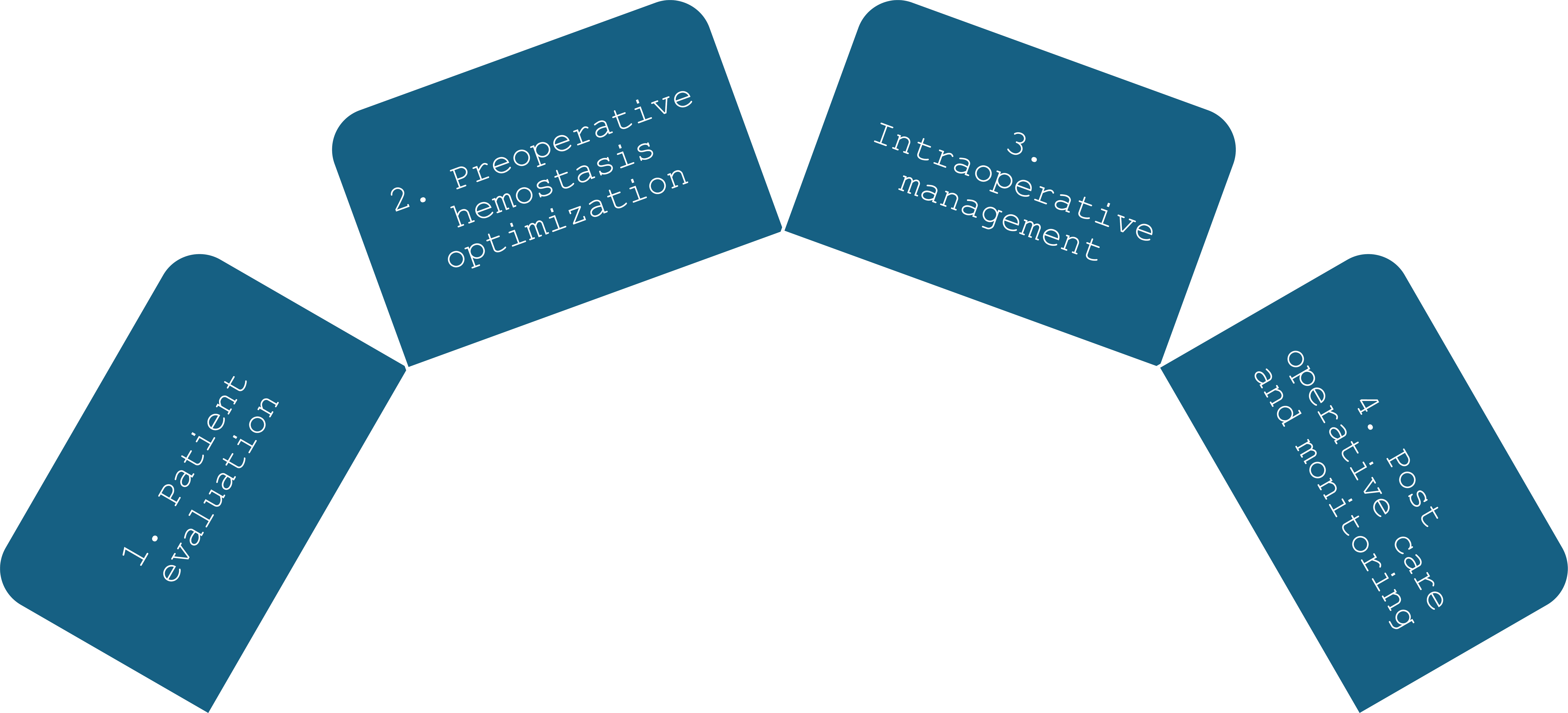
Haemophilia management in dental practice.
1.6. Laboratory Investigations
Laboratory investigations are crucial in diagnosing, managing, and monitoring haemophilia (Table 1). The primary goal is to assess the levels of specific clotting factors, which directly influence treatment strategies [22, 23].
| Investigation | Purpose |
|---|---|
| Activated Partial Thromboplastin Time (aPTT) | Prolonged in haemophilia due to intrinsic pathway deficiency; used for initial screening |
| Prothrombin Time (PT) | Normal in haemophilia; differentiates from other coagulation disorders |
| Factor Assays (FVIII, FIX) | Measures factor VIII (FVIII) or factor IX (FIX) levels to confirm diagnosis and severity |
| Inhibitor Testing (Bethesda Assay) | Detects inhibitors that neutralize factor replacement therapy; critical for treatment planning |
| Thrombin Generation Tests | Assesses thrombin generation and efficacy of treatment, including non-factor therapies |
| Thromboelastography (TEG) | Evaluates the viscoelastic properties of blood to monitor clot formation and stability |
| Genetic Testing | Identifies mutations in F8 (haemophilia A) or F9 (haemophilia B) genes; aids in diagnosis and genetic counseling |
1.6.1. Coagulation Assays
The initial step involves performing standard coagulation assays, including activated partial thromboplastin time (aPTT) and prothrombin time (PT). In patients, aPTT is typically prolonged due to the involvement of the intrinsic pathway, while PT remains normal [22, 23]. The aPTT test is susceptible to deficiencies in FVIII and FIX, making it a valuable tool for initial screening [22].
1.6.2. Factor Assays
Once prolonged aPTT is identified, specific factor assays quantify factor levels to confirm diagnosis and determine severity [22].
1.6.3. Inhibitor Testing
Inhibitors that neutralize replacement factors complicate treatment, making routine testing essential for patients on factor replacement therapy [22, 23]. The Bethesda assay quantifies inhibitor levels to guide treatment.
1.6.4. Thrombin Generation Tests
Thrombin generation tests provide insights into hemostatic potential and help evaluate treatment efficacy, particularly for non-factor therapies such as emicizumab [24]. Thromboelastography assesses blood’s viscoelastic properties and provides real-time data on clot formation [24].
1.7. Preoperative Hemostasis Optimization
Preoperative hemostasis optimization is critical for patients with haemophilia undergoing surgery.
1.8. Factor Replacement Therapy
Factor replacement therapy is essential before surgery to raise plasma levels of the missing clotting factor and prevent bleeding . Factor concentrates are given shortly before surgery to ensure peak levels during the procedure. WFH guidelines recommend a loading dose within three days before surgery, with preoperative and postoperative monitoring to ensure adequate hemostasis [25].
1.9. Recombinant Factors
Advances in recombinant factor therapies have enhanced treatment safety and effectiveness. Recombinant factor VIIa can be used to develop inhibitors, providing an alternative approach to restoring hemostasis.
1.10. Antifibrinolytics
Antifibrinolytic agents (TXA and EACA) are lysine analogs that inhibit plasminogen activation, preventing fibrin clot breakdown and maintaining hemostasis [25, 26]. Systematic reviews show that they significantly decrease hemorrhage [27].WFH recommends combining antifibrinolytics with factor replacement therapy. TXA should be given preoperatively and continued briefly postoperatively [26].
1.11. Intraoperative Management of Haemophilia
Intraoperative management is critical to minimize the risk of bleeding during surgical procedures. It involves several key considerations, including anesthesia techniques, local hemostatic measures, minimizing trauma, and pharmacological support.
1.12. Anesthesia Considerations
Anesthesia should avoid techniques that could cause significant bleeding. Nerve blocks in vascular areas should be avoided due to hematoma risk [28]. General or local anesthesia with careful technique minimizes tissue trauma [29]. The anesthesiology team must ensure appropriate factor replacement therapy before the procedure [30].
1.13. Local Hemostatic Measures
1.13.1. Pressure Application and Sutures
To control bleeding, direct pressure should be applied to any bleeding site immediately after surgical manipulation [31]. Proper suturing techniques are crucial for securing tissues and minimizing dead space, thereby preventing hematoma formation. Absorbable sutures may be beneficial in certain situations to reduce the need for suture removal and minimize trauma [31].
1.13.2. Local Hemostatic Agents
Local hemostatic agents can be classified into two categories. Passive hemostatic agents, such as collagen-based, cellulose-based, and gelatin-based products, primarily function mechanically by providing a scaffold for platelet aggregation, thereby promoting clot formation. Active hemostatic agents, including thrombin and fibrin sealants, directly influence the coagulation cascade to enhance hemostasis [28].
1.13.3. Collagen-Based Hemostatic Agents
Collagen-based agents (Avitene, Helistat) utilize purified bovine collagen to stimulate platelet aggregation and control bleeding. They are effective in moderate to severe bleeding and are absorbed within 10–14 days; however, pH sensitivity can compromise efficacy. Helistat is contraindicated in infected sites [30, 31].
1.13.4. Cellulose-Based Hemostatic Agents
Surgicel, an oxidized regenerated cellulose, acts as an absorbable matrix that expands significantly upon contact with blood, enhancing hemostasis through mechanical pressure. This agent is noted for its bacteriostatic properties and is effective in controlling minor bleeding, although it may delay bone regeneration. Other cellulose-based products, such as Actcel and Gelitacel, enhance platelet aggregation and physically stabilize clots, demonstrating hypoallergenic and bacteriostatic properties [30]. These agents are beneficial in surgical settings where rapid hemostasis is required; however, they require careful consideration regarding their impact on wound healing and potential complications associated with retention.
1.13.5. Gelatin-Based Hemostatic Agents
Gelatin-based products (Gelfoam) absorb blood and expand to facilitate hemostasis, with minimal tissue reaction and absorption within 4–6 weeks. Studies show effectiveness when combined with thrombin in complex bleeding scenarios. Gelatin’s biocompatibility makes it a preferred choice in many surgical contexts.
1.13.6. Active Hemostatic Agents
Active hemostatic agents, such as thrombin and fibrin sealants like Tisseel, directly interact with the coagulation cascade. Thrombin converts fibrinogen to fibrin, effectively sealing bleeding sites. Fibrin sealants offer hemostatic properties and promote tissue healing and angiogenesis, making them valuable in complex surgical procedures such as bone grafting. However, their use is limited in cases of vigorous bleeding, necessitating careful evaluation of the bleeding source and the type of hemostatic agent employed [28]. Selecting appropriate local hemostatic agents is critical, as each type offers unique benefits and limitations. Understanding the mechanisms, absorption rates, and potential complications associated with these agents is essential for optimizing patient outcomes during surgical interventions (Table 2).
| Local Hemostatic Agents | Local Hemostatic Agents | Local Hemostatic Agents | Local Hemostatic Agents |
|---|---|---|---|
| Passive hemostatic agents | Source | Action | Remarks |
| Collagen Based | |||
| Micro fibular collagen Avitene, Colla-plug, Colla-tape, Colla-cote | Purified bovine collagen | • Stimulates aggregation of platelets to form a thrombus | • . Thrombin is not effective with this product because of the pH • Adsorbed in 10-14 days • Effective in moderate to severe capillary, venous, or minor arterial bleeding |
|
Absorbable collagen hemostatic sponge Helistat |
Freeze-dried bovine tendon | • Causes aggregation of platelets • Mechanical obstruction to bleeding • Forms a three-dimensional matrix, strengthening the clot |
• Promotes wound protection and control of oozing and bleeding from clean oral wounds • Contraindicated in infected or contaminated wounds |
| Cellulose-based | |||
| Surgicel | Oxidized regenerated plant-based cellulose | • Absorbable physical matrix for clotting initiation • Expands 7-10times • Hemostasis by mechanical pressure |
• Thrombin is not effective with this product because of the pH • More bacteriostatic than other agents • Used in dry form • Adsorption in 4-8 weeks • May delay bone regeneration |
| Actcel, Gelitacel | Treated, sterilized cellulose mesh | • Acts biochemically on intrinsic pathways, enhancing platelet aggregation • Physically aids in clot stabilization • Expands 3-4 times |
• Dissolve in 1-2 weeks • Does not affect wound healing • Hypoallergenic • Bacteriostatic • Gelitacel absorbs as fast as 4 days |
| Gelatin based | |||
| Gelfoam | Purified pork skin gelatin | • Absorbs 40 times its weight • Expands 2 times the volume • Provides a clotting framework |
• Little tissue reaction • Absorbs in 4-6 weeks • Do not use on contaminated or infected wounds |
| Active hemostatic agents | |||
| Thrombin | Bovine plasma, human plasma, or recombinant DNA | • Converts fibrinogen to fibrin | • Used as a dry powder, solution mixed with gelatin sponge, or as a spray • Used to treat moderate to severe bleeding • Avoid injection into the bloodstream or use near large open vessels. |
| FioSeal | Bovine-derived gelatin granules coated in human-derived thrombin | • Swells in 10%-20% • Mechanically seals the bleeding site • Activates the common pathway of the coagulation cascade |
• Resorption in 6-8 weeks • It can be used in irregular spaces • Effective in hard and soft tissues |
| Sealants | |||
| Fibrin sealants | |||
| Tisseel | Natural, synthetic combination of hemostatic agents and tissue adhesive | • Forms a barrier impervious to the flow of most liquids • Impacts tissue angiogenesis and wound healing |
• It can be used in bone grafting, especially in sinus lift surgery • It can be used in patients with insufficient fibrinogen • It can be used on patients receiving heparin • Not effective in vigorous bleeding |
| Newer hemostatic agents | |||
| Chitosan-based | |||
| HemCon | Positively charged polysaccharides derived from crustacean shells | • Attracts negatively charged RBCs, forming a cellular lattice that forms an artificial clot | • The clot formed is independent of intrinsic or extrinsic pathways • Effective for patients on anticoagulation medication • Does not cause adverse reactions in shellfish-sensitive patients |
| Hemostatic Solutions | |||
| Tannic acid | Like plant polyphenol tannin | • Causes local vasoconstriction | |
| Passive hemostatic agents | |||
| Tranexamic acids | • Inhibits plasminogen, stabilizes the clot | • It can be used as a perioperative mouthwash or rinse • Helpful in minor oral surgical procedures in patients with bleeding diathesis or who are on anticoagulant medication |
|
| Epsilon aminocaproic acid | Less potent than tranexamic acid | ||
| Hemocoagulase botroclot | Derived from snake venom | • Accelerates conversion of prothrombin to thrombin • Causes the transformation of fibrinogen to fibrin monomer |
• Contraindicated in conditions with risks of thrombosis formation |
| Bone hemostats | |||
| Bone wax | Water-insoluble beeswax, wax, paraffin, and softening agents | • Physically, tamponades localized medullary bone bleeding | • Non-resorbable • Interferes with localized bone healing • Use with caution in future bone graft sites |
| Ostene | Water-soluble alkylene oxide copolymer | • Its use is similar to bone wax. | • Eliminated from the body in 48 hours • It does not cause infections, inflammatory reactions, or interference with bone healing. |
1.14. Pharmacological Support
Use of Local Vasoconstrictors: Local vasoconstrictors, such as epinephrine, can be used in conjunction with local anesthetics to reduce blood flow to the surgical site. However, caution should be exercised to avoid excessive vasoconstriction, which could compromise tissue perfusion.
1.14.1. Hemostatic Agents
Pharmacological agents, such as tranexamic acid, may be administered preoperatively or intraoperatively to enhance hemostasis [30, 31]. Anticoagulants are useful in managing thromboembolic disorders, and their use requires careful consideration of their mechanisms of action, renal function, and discontinuation guidelines before surgical procedures.
- Warfarin is a vitamin K antagonist that inhibits the synthesis of clotting factors II, VII, IX, and X, which are essential for the coagulation cascade. Its half-life ranges from 20 to 60 hours, necessitating careful monitoring of renal function, particularly creatinine clearance. For patients with a creatinine clearance greater than 80 ml/min, it is recommended to hold warfarin for 48 hours before surgery. For individuals with a clearance between 50–80 ml/min, a 72-hour hold is recommended. Discontinuation is based on the International Normalized Ratio (INR), with surgery contraindicated if the INR exceeds 3.5 [32].
- Dabigatran is a direct thrombin inhibitor that inhibits both free and fibrin-bound thrombin, thus preventing the conversion of fibrinogen to fibrin. Its half-life is approximately 12 to 17 hours and does not require INR monitoring. Patients with a creatinine clearance of 80 ml/min or higher do not typically need to discontinue before dental procedures. However, it is advisable to consult a physician if more than two teeth are to be removed.
- Rivaroxaban and Apixaban are selective factor Xa inhibitors with half-lives of 9–13 and 9–14 hours, respectively. They do not require INR monitoring and are generally not discontinued for dental extractions unless more than 2–3 teeth are involved. Renal clearance thresholds are ≥30 ml/min (rivaroxaban) and ≥15 ml/min (apixaban).
- Low-molecular-weight heparins (LMWHs), such as enoxaparin and dalteparin, inhibit factor Xa and thrombin. Their half-lives are approximately 4.5 hours and 2.2 hours, respectively. These agents are often used for bridging therapy in patients at risk for thromboembolic events during the perioperative period. The management of LMWH requires careful timing of administration relative to surgical procedures to minimize bleeding risks while ensuring adequate anticoagulation.
- Antiplatelet agents such as acetylsalicylic acid (aspirin), clopidogrel, and ticagrelor inhibit platelet aggregation. Aspirin has a short half-life of 15–20 minutes and may need to be discontinued based on dosing and the specific surgical procedure. Clopidogrel, with a half-life of 7–9 hours, typically requires discontinuation 5–7 days before elective surgeries, while ticagrelor does not need to be stopped for elective surgeries unless the bleeding risk is high. The timing of discontinuation for these agents often depends on the procedure's bleeding risk and the patient's overall thromboembolic risk profile.
Managing anticoagulants and antiplatelet medications in the perioperative setting is complex and requires a thorough understanding of each agent's pharmacokinetics, renal function considerations, and the specific surgical context. Clinicians must balance the risks of thromboembolism against the potential for bleeding complications, making individualized patient assessments critical for optimal outcomes.
1.15. Minimizing Trauma
Surgical techniques should prioritize gentle tissue handling and be minimally invasive [29]. This approach reduces trauma, shortens recovery time, and minimizes the risk of complications [29, 31].
1.16. Postoperative Care for Hemophilic Patients
Postoperative care is critical to ensure optimal recovery and minimize the risk of complications. This care involves several key components, including hemostasis monitoring, appropriate medication management, patient instructions for postoperative care, and emergency protocols for managing delayed bleeding.
1.16.1. Hemostasis Monitoring
1.16.1.1. Continuous Observation for Signs of Bleeding:
Patients require vigilant monitoring for excessive bruising, swelling, or hematoma formation at the surgical site, as well as vital signs for internal bleeding. Signs of bleeding should prompt immediate intervention, including potential factor replacement therapy.
1.16.1.2. Prescribing Analgesics
Acetaminophen is preferred over NSAIDs, which inhibit platelet function and increase bleeding risk. Opioids may be considered for severe pain but require careful monitoring [32].
1.16.2. Patient Instructions
1.16.2.1. Postoperative Care Tips
Patients should receive clear instructions to minimize complications during recovery. Recommendations may include:
- To avoid trauma to the oral cavity, a softer diet is preferably advised [14].
- Patients should be instructed to avoid spitting, which can create negative pressure in the oral cavity and further complicate the situation.
- Limit physical activity and avoid strenuous movements that could stress the surgical site and provoke bleeding [32].
1.17. Advances and Updates in Management
Recent advancements aim to improve treatment adherence, reduce infusion frequency, and enhance patients' overall quality of life. The introduction of long-acting recombinant factor products has reduced the frequency of spontaneous bleeds [33, 34]. However, the presence of inhibitor antibodies that neutralize the effectiveness of factor replacement therapy complicates treatment and can lead to increased morbidity and mortality in affected patients [34].
1.18. Extended Half-Life Clotting Factors
Recombinant factor VIII was the first to be developed, followed by the successful replication of factor IX. Recombinant factor VIII products can be categorized by their production method: animal or human cell culture. Initial products used animal proteins along with human serum albumin, whereas newer drugs use human-origin proteins in a culture medium without albumin. Modern protein production has successfully eliminated the addition of either animal or human proteins in its ingredients. One key area of experimentation has been modeling recombinant proteins with extended half-lives. This is because the required infusion time to sustain clotting factor levels greater than 1% is quite long: approximately 12 hours for factor VIII and 16–18 hours for factor IX [35]. This places an additional burden on patients, particularly children and adults, while simultaneously increasing the cost of the procedure. To address this problem, modifying coagulation factors, such as PEGylation or cross-linking with the Fc fragment of antibodies, has been proposed [36]. Extended half-life recombinant FVIII and FIX concentrates have been developed to prolong the duration of action and allow less frequent dosing than traditional factor replacement therapies, thereby significantly reducing the treatment burden [36]. These factors have shown promising results in clinical trials, demonstrating effective hemostatic control with fewer infusions per week, which is beneficial for patients with severe haemophilia who require prophylactic treatment to prevent bleeding episodes [36]. This has also raised awareness of the potential discordance between one-stage activated partial thromboplastin time (aPTT)-based assays and chromogenic factor assays, necessitating careful monitoring and adjustment of treatment protocols [35].
1.19. Non-replacement Therapy for Haemophilia
Antibody-based therapies offer an alternative to EHL drugs by reducing injection frequency. Emicizumab, a humanized bispecific monoclonal antibody (IgG4), mimics factor VIII activity by binding factors IX and X, accelerating their activation. Concizumab, a humanized IgG4 antibody binding the Kunitz-2 domain, modulates thrombin generation and induces procoagulant effects [30, 37].
1.20. Gene Therapy
Gene therapy addresses the underlying genetic defect by delivering a functional copy of the deficient gene, enabling endogenous production of the deficient clotting factor [30]. Early clinical trials show sustained increases in factor levels, reducing bleeding episodes and the need for factor replacement [30]. Adeno-associated virus (AAV) vectors are commonly used for gene delivery. Challenges remain for patients with inhibitors, who have been excluded from studies [30]. Ongoing research focuses on optimizing delivery methods for long-term safety and efficacy [37].
1.21. Treatment of Haemophilia using Blood-purified Factors VIII and IX
In the 1950s and 1960s, whole blood plasma transfusions were the prevailing mode of treatment for haemophilia until cryoprecipitation technology emerged in the second half of the 1960s, enabling the precipitation of necessary blood proteins under a specific thawing regimen for fresh-frozen plasma. In this manner, it is feasible to obtain concentrated clotting factors, thereby significantly reducing the infusion volume.
1.22. Cellular Therapy of Haemophilia
Patients undergoing cell therapy have their endothelial progenitor cells and stem cells transformed in vitro to express coagulation factors before transplantation. Numerous studies have demonstrated successful transplantation of transformed hematopoietic stem cells expressing FVIII or FIX using retroviral or lentiviral vectors. Research on induced pluripotent stem cells (iPSCs) for haemophilia treatment has also been conducted, as the pool of cells expressing coagulation factors is larger [38].
1.23. Role of Dental Lasers in Minimizing Bleeding
Dental lasers minimize bleeding by precisely targeting soft tissues and promoting coagulation while minimizing surrounding damage. Studies show diode lasers significantly decrease intraoperative bleeding and postoperative discomfort, contributing to quicker recovery and less pain [39].
1.24. Updated Guidelines and Consensus Statements
Recent guidelines emphasize individualized treatment plans considering patient needs, including EHL factors and gene therapy where appropriate [35, 38, 39].
1.25. Interdisciplinary Collaboration
Interdisciplinary collaboration is essential; the involvement of various healthcare professionals ensures that patients receive holistic and coordinated care [39]. The management often necessitates surgical interventions, which require careful perioperative planning. Integrating different specialties enables a more comprehensive assessment of the patient's health status and facilitates the development of individualized treatment plans that address both medical and psychosocial aspects of care. Collaborative efforts enhance patient education and improve adherence to treatment regimens [34, 40].
1.26. Challenges in Interdisciplinary Collaboration
Despite the recognized benefits of interdisciplinary collaboration, several challenges persist. One significant barrier is the lack of awareness and understanding of haemophilia among healthcare providers outside specialized centers. This can lead to inconsistent care and management practices, particularly in emergencies requiring immediate intervention [40]. Additionally, communication gaps across specialties can hinder the timely exchange of critical information in patient care. Another challenge is the need for ongoing education and training for healthcare professionals who care for patients with haemophilia. Continuous professional development ensures that all team members are up to date with the latest treatment protocols and management strategies [39]. Patients can receive comprehensive care through a team-based approach that involves multiple healthcare professionals.
1.27. Clinical Perspective: What Dentists Need to Know Today?
With the advent of extended half-life clotting factors, non-factor therapies such as emicizumab, and gene therapy trials, the dental management of patients with haemophilia is evolving. For dentists, this means risk assessment must include not only bleeding history but also the specific therapy regimen, since newer agents may reduce spontaneous bleeding while altering the response to factor replacement in cases of surgical bleeding. Coordination with hematologists is critical to understand whether the patient is on prophylaxis, requires factor supplementation, or is receiving non-factor therapy that modifies coagulation pathways. In preoperative planning, dentists should adopt a tailored approach. Postoperatively, these therapies may reduce the duration of bleeding risk, but vigilant monitoring and patient education remain essential, particularly for invasive procedures. Table 3 presents a dental management quick clinical decision table for haemophilia patients.
| I. Pre-treatment Assessment | |
|---|---|
| Step | Clinical decision |
| Type of haemophilia | A (Factor VIII) / B (Factor IX) |
| Severity | Mild (>5%), Moderate (1–5%), Severe (<1%) |
| History | Previous bleeding episodes, inhibitors |
| Physician consult | Mandatory before invasive procedures |
| Baseline labs | aPTT, factor level (if advised) |
| II. Dental Procedures & Management | |
| Dental Procedure | Management Decision |
| Examination, radiographs | No factor replacement needed |
| Oral prophylaxis | Usually safe; avoid trauma |
| Local anesthesia (infiltration) | Safe |
| Inferior alveolar nerve block | Avoid unless factor coverage. |
| Simple extraction | Factor replacement ± local hemostatic |
| Surgical extraction | Factor replacement + hospital setting |
| Pulp therapy (primary teeth) | Prefer non-surgical approaches |
| Scaling (deep) | Factor coverage if bleeding is expected |
| III. Local Hemostatic Measures (Always Use) | |
| Method | Example |
| Pressure | Gauze pack |
| Sutures | Resorbable sutures |
| Topical agents | Oxidized cellulose, gelatin sponge |
| Antifibrinolytic | Tranexamic acid mouthwash |
| IV. Factor Replacement Guide (General) | |
| Severity | Factor Coverage |
| Mild | Often not required |
| Moderate | Required for invasive procedures |
| Severe | Mandatory for any surgical procedure |
| - | The hematologist decides the exact dose. |
| V. Drug Prescription Rules [High-Yield] | |
| Drug Type | Recommendation |
| Analgesic | Paracetamol |
| Avoid | Aspirin, NSAIDs |
| Antibiotics | Safe as per the indication |
| VI. Post-operative Instructions | |
| Instruction | Purpose |
| Soft diet | Prevent trauma |
| Avoid rinsing for 24 hrs. | Clot stability |
| Tranexamic mouthwash | Prevent clot breakdown |
| Emergency contact | Early bleeding management |
| VII. Pediatric Dentistry–Specific Points | |
| Aspect | Decision |
| Emphasis | Prevention over intervention |
| Fluoride therapy | Strongly recommended |
| Sealants | Safe |
| Extraction planning | Prefer referral/hospital setup. |
| Behavior management | Avoid techniques causing trauma. |
CONCLUSION
This narrative review does not follow a systematic methodology, which is a limitation. Haemophilia requires careful management to mitigate bleeding complications. The evolution of treatment strategies, including prophylactic factor replacement, inhibitor management, and EHL therapies, improves patient outcomes and quality of life. Dental care is paramount; increased awareness, education, and collaboration among healthcare providers are essential for improving dental health outcomes for hemophilic patients.
AUTHORS’ CONTRIBUTIONS
The authors confirm their contributions to the paper as follows: S.K.S. and A.K.: Study conception and design; S.K.S. and A.V.: Drafting of the manuscript; A.S. and M.J.: Critically evaluated the manuscript. All authors reviewed the results and approved the final version of the manuscript.
LIST OF ABBREVIATIONS
| aPTT | = activated partial thromboplastin time |
| PT | = Prothrombin Time |
| TMJ | = Temporomandibular Joint |
| WFH | = World Federation of haemophilia |
| DeBRATT | = Dental Bleeding Risk Assessment and Treatment Tool |
| TXA | = Tranexamic acid |
| EACA | = Epsilon aminocaproic acid |
| LMWH | = Low Molecular weight heparin |
| NSAID | = Non-steroidal anti-inflammatory drugs |
| AAV | = Adeno-associated virus |
| iPSC | = Induced pluripotent stem cells |
ACKNOWLEDGEMENTS
Declared none.

